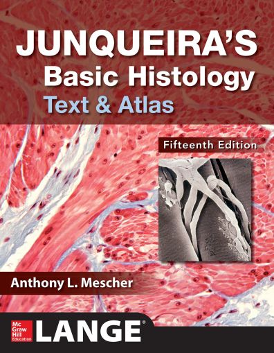
Junqueira's Basic Histology: Text And Atlas, 15th Edition
-
Код:KS2771
Junqueira's is written specifically for students of medicine and other health-related professions, as well as for advanced undergraduate courses in tissue biology - and there is nothing else like it
This trusted classic delivers a well-organized and concise presentation of cell biology and histology that integrates the material with that of biochemistry, immunology, endocrinology, and physiology, and provides an excellent foundation for subsequent studies in pathology.
With this 15th edition, Junqueira’s Basic Histology continues as the preeminent source of concise yet thorough information on human tissue structure and function. For over 45 years this educational resource has met the needs of learners for a well-organized and concise presentation of cell biology and histology that integrates the material with that of biochemistry, immunology, endocrinology, and physiology and provides an excellent foundation for subsequent studies in pathology. The text is prepared specifically for students of medicine and other health-related professions, as well as for advanced undergraduate courses in tissue biology. As a result of its value and appeal to students and instructors alike, Junqueira’s Basic Histology has been translated into a dozen different languages and is used by medical students throughout the world.
Unlike other histology texts and atlases, the present work again includes with each chapter a set of multiple-choice Self-Test Questions that allow readers to assess their comprehension and knowledge of important points in that chapter. At least a few questions in each set utilize clinical vignettes or cases to provide context for framing the medical relevance of concepts in basic science, as recommended by the US National Board of Medical Examiners. As with the last edition, each chapter also includes a Summary of Key Points designed to guide the students concerning what is clearly important and what is less so. Summary Tables in each chapter organize and condense important information, further facilitating efficient learning.
Each chapter has been revised and shortened, while coverage of specific topics has been expanded and updated as needed. Study is facilitated by modern page design. Inserted throughout each chapter are more numerous, short paragraphs that indicate how the information presented can be used medically and which emphasize the foundational relevance of the material learned.
The art and other figures are presented in each chapter, with the goal to simplify learning and integration with related material. The McGraw-Hill medical illustrations, now used throughout the text, are the most useful, thorough, and attractive of any similar medical textbook. Electron and light micrographs have been replaced throughout the book as needed, and they again make up a complete atlas of cell, tissue, and organ structures fully compatible with the students’ own collection of glass or digital slides. Health science students whose medical library offers AccessMedicine among its electronic resources (which includes more than 95% of U.S. medical schools) can access a complete human histology Laboratory Guide linked to the virtual microscope at the URL given below. This digital Laboratory Guide, which is new with this edition of the text and unique among learning resources offered by any histology text and atlas, provides both links to the appropriate microscope slides needed for each topic and links to the correlated figures or tables in the text. Those without AccessMedicine will lack the digital Laboratory Guide, but may still study and utilize the 150 virtual microscope slides of all human tissues and organs, which are available at: http://medsci.indiana.edu/junqueira/virtual/junqueira.htm.
As with the previous edition, the book facilitates learning by its organization:
-
An opening chapter reviews the histological techniques that allow understanding of cell and tissue structure.
-
Two chapters then summarize the structural and functional organization of human cell biology, presenting the cytoplasm and nucleus separately.
-
The next seven chapters cover the four basic tissues that make up our organs: epithelia, connective tissue (and its major sub-types), nervous tissue, and muscle.
-
Remaining chapters explain the organization and functional significance of these tissues in each of the body’s organ systems, closing with up-to-date consideration of cells in the eye and ear.
With its proven strengths and the addition of new features, I am confident that Junqueira’s Basic Histology will continue as one of the most valuable and most widely read educational resources in histology. Users are invited to provide feedback to the author with regard to any aspect of the book’s features.
Anthony L. Mescher
Indiana University School of Medicine
Contens:


